
HUMAN EYE: ANATOMY AND STRUCTURE
 The eye is an extremely delicate and complex organ.
To simplify matters, we can compare it to a camera with an auto-focus lens.
A camera lens is convex, making luminous rays converge and then focusing them on film. In an eye, there are two lenses, the outermost is the cornea and the innermost the crystal.
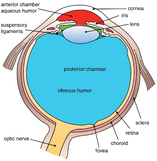
Diaphragm: this mechanism in a camera can change its diameter to let more or less light enter and make a better impression on film. The iris is the part of the diaphragm which gives color to the eye.
Film: this part of a camera is exposed to light and when developed gives photographs. In the eye the retina changes light into electric impulses that move from the optical nerve to the brain.
Another essential aspect of photography is correct focus. By moving the lens forward or backward with respect to the film the photo comes into focus. In an eye, depth, shape and position of the crystal are modified by a muscle through accommodation. Since accommodation diminishes progressively through the years, eyeglasses need to be used after age forty. Whether a crystal is replaced to eliminate a cataract or for optical reasons, it loses this ability and the eye becomes presbiopic.
The cornea and crystal lenses make up the eye’s optical system.
The cornea has about 43 positive dioptres while the crystal has 20.
These two lenses focus external images on the retina and on the macula or center of the retina.
Cornea
The cornea is made up of transparent proteins and is between 500 central thousands of millimeters to 900 outer millesimi deep. This is the eye’s strongest lens where the most popular techniques of refractive surgery are practiced.
Crystal
The crystal is the eye’s internal lens located behind the iris. It allows us to see things clearly both near and far until presbyopia takes over. When it is opaque there is a problem with cataracts.
Vitreous
This elastic, transparent, filamentous gelatin fills the eye giving it the necessary pressure to maintain its spherical shape. Since it is made up almost entirely of water, it allows the retina to stay attached to the external layers.
Iris
This circular muscular membrane is behind the cornea and in front of the crystal. It has a central hole (the pupil) and has different colors which color the eyes.
Pupil
This is the circular hole in the iris that can change size so that more or less light enters the eye.
Retina
This is the eye’s nervous structure that covers almost the entire internal surface. It is made up of many different cells on different layers. The most important central part is called “macula”. There are three million cells called cones in the macula that give clear vision of objects. In the outermost part of the retina another 70-80 million cells called rods give night vision and groups things together.
Optic Nerve
One million retinal cell fibers make up the cable which connects the eye to the brain called optic nerve. A lesion of the optic nerve can therefore give eyesight problems even in an otherwise perfect eye. It can be damaged through an increase in internal eye pressure (glaucoma).
.